

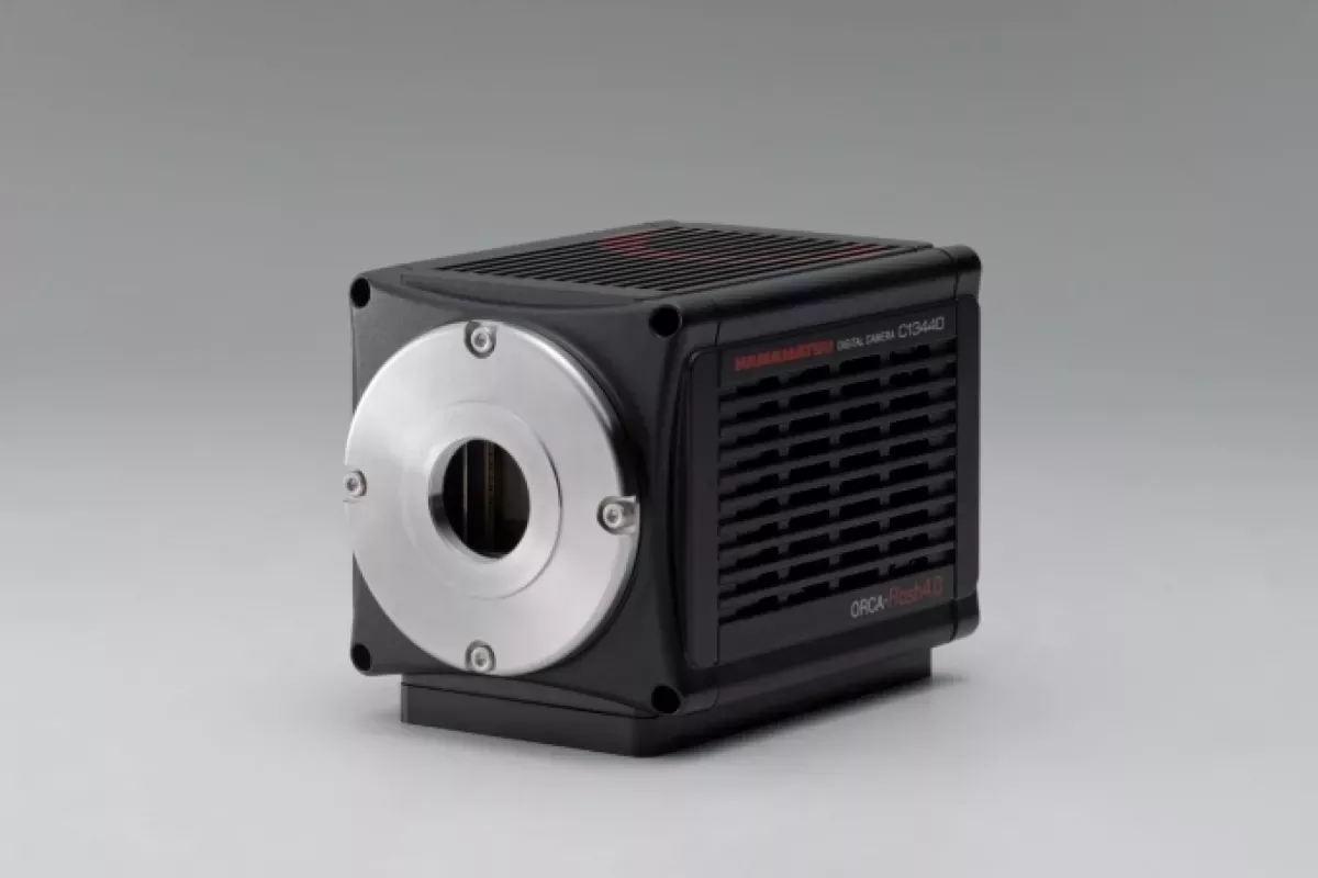
Camera ORCA Flash4.0 v3r Hamamatsu
The ORCA-Flash 4.0 v3 camera from Hamamatsu is designed for high-speed, high-sensitivity scientific imaging. It offers 4 MPixel resolution with 6.5 µm pixels and a large field of view. It can capture up to 100 frames/s at full resolution and up to 25,655 frames/s in lightsheet mode, thanks to ROI-optimised readout. Its quantum efficiency reaches 82% at 560 nm, with very high sensitivity between 400 and 900 nm. The camera has very low read noise (1 e- median at 40 fps) and a background noise of 0.06 e-/pixel/s. Digitisation is possible in 8, 12 or 16 bits. It offers different readout modes: normal, lightsheet and W-View, offering maximum flexibility in terms of regions of interest and exposure times.
ORCA Flash4.0 v3r Hamamatsu camera features
- Resolution: 4 MPixels, 6.5 µm² pixels
- Field of view: x3
- Acquisition speed: 100 to 25,655 fps depending on ROI
- ROI 2048 x 2048: 100 fps
- ROI 2048 x 8: 25,655 fps
- Quantum efficiency: 82% at 560 nm
- Sensitivity: 400-900 nm
- Median read noise: 1 e- @ 40 fps
- Background noise: 0.06 e-/pixel/s
- Digitisation: 8, 12 or 16 bits
- Readout modes: normal, lightsheet, W-View
- Independent readout for regions of interest and exposure directions
Applications
- Time lapse with wide field of view
- Very fast calcium imaging
- Rtiometric imaging
- Light sheet microscopy