

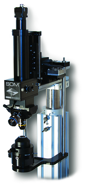
SOM Simple Moving Microscope SUTTER INSTRUENT
Sutter Instrument’s SOM (Son of MOM®) open stand microscope is a compact and simple platform dedicated to in vitro and in vivo experiments, offering robotic positioning of the objective via a motorised manipulator (MP-285/MPC-385).
Inspired by the MOM® microscope, it maximises the space under the objective, essential for patch-clamp or multi-cell applications, while using Multi-Link™ software to automatically coordinate the positioning of recording and stimulation pipettes.
The IR LED OCC condenser and configurable excitation port ensure optimal illumination and adaptive flexibility.
Features of the SOM open stand microscope
- Based on an MP-285 or MPC-385 motorised manipulator, controlled X, Y, Z
- Allows rapid robotic positioning of the objective (no heavy motorised stage)
- Ideal for switching between in vivo and in vitro without reconfiguration
- Transmitted IR illumination + epifluorescent fluorescence via 2-position filter cube
- C-mount excitation port with cage assembly for additional sources
- Mobile IR LED OCC condenser synchronised with microscope movement
- Free Multi-Link™ software for synchronised pipette and objective positioning
Technical specifications of the SOM open stand microscope
| Caractéristique | Valeur |
|---|---|
| Positioning mode | X, Y, Z robotics via MP-285/MPC-385 |
| Mobile lens | Can be moved without a heavy turntable |
| Illumination | Transmitted IR (LED + IR-compatible CCD), epifluorescence, optional OCC condenser |
| Filter cube | 2 positions, one IR passage possible |
| Excitement port | C-mount thread + adaptable cage holes for multiple sources |
| Software | Multi-Link™ (free), integrates motion manipulator and lenses |
| Compatible manipulator | MP-285, MP-225, MP-385, MPC-200 (USB controller) |
| Versions | SOM-T (MT-75T), SOM-XT (MT-75XT), MP-225, MP-285 or 4-axis QUAD configurations |
| Options | OCC LED condenser, CAM, camera, TLED+ white illumination |
Applications
- In vivo and in vitro patch-clamp experiments with optimised positioning
- Transmitted IR and fluorescence imaging on live cells or slices
- Photostimulation via adaptable excitation port
- Multi-cell, multi-pipette workflow thanks to robotic coupling
Documentation
SOM microscope flyer
PDF file – 658.5 ko