

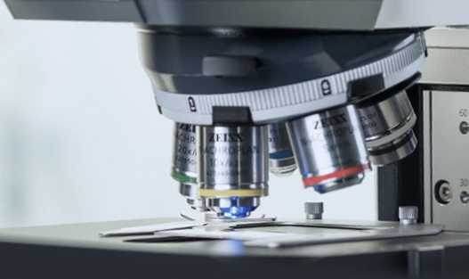
Camera CellCam Kikker Cairn Research
The CellCam Kikker is a scientific CMOS (sCMOS) camera from Cairn Research with high sensitivity and a very large field of view, suitable for fluorescence imaging of living cells, brightfield, phase contrast and DIC. It is thermally cooled to minimise dark current, has a quantum efficiency of around 85%, and supports 12-bit digitisation (or 16-bit depending on configuration) to optimise low-light and high-dynamic performance.
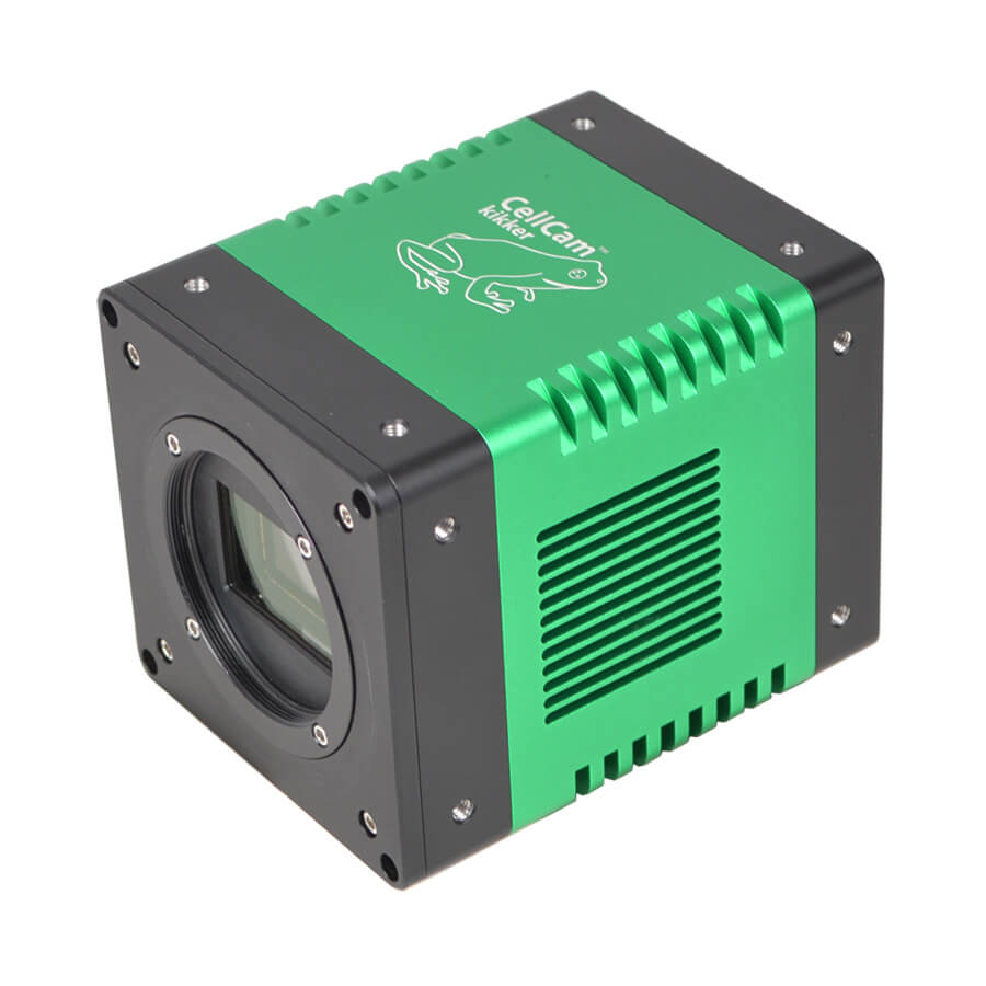
CellCam Kikker Cairn Research Camera features
- Thermoelectric cooling for low dark current
- Quantum efficiency ≈ 85% (peak around 530 nm), optimised for sensitive low-signal imaging
- 12-bit (USB) or 16-bit (CoaXPress) digitisation for wide dynamic range
- High resolution flexibility: 11.7 MP default (bin 2×2), up to 46.8 MP with software unlocking
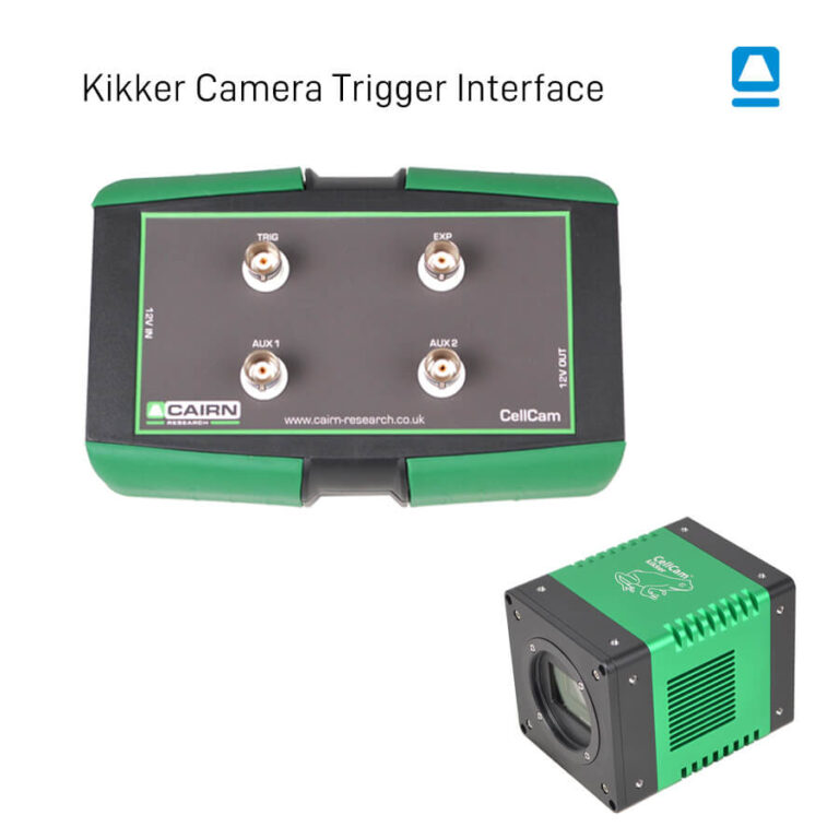
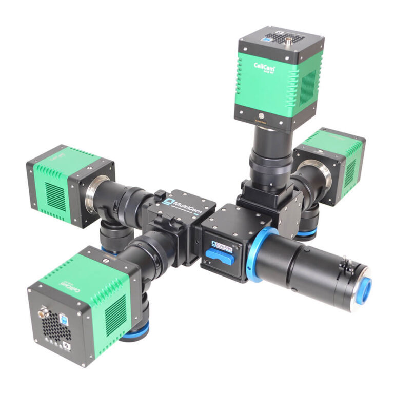
- Multi-interface: standard USB 3.0 Type B + CoaXPress for fast frame rates and reliable synchronisation
- C-Mount included (M48×0.75) or T2 (M42×0.75 depending on configuration) with adapter supplied
Technical specifications of the Camera CellCam Kikker Cairn Research
| Caractéristique | Valeur |
|---|---|
| Sensor | sCMOS, backlit, mono |
| Native resolution | 2304 × 2304 pixels (≈11.7 MP) |
| Pixel size | 4.63 µm (2.315 µm in high-resolution mode) |
| Active sensor zone | ~13 mm × 19 mm (~23 mm diagonal) |
| Quantum efficiency | ≈ 85% (≈ 90% according to some sources) |
| Playback noise | ~0.7 e- RMS |
| Depth of well | ~15,000 electrons |
| Maximum frequency | ~89 fps in CoaXPress mode |
| Scanning | 12-bit (USB) or 16-bit (CoaXPress) |
| Cooling | Peltier (≈ -30 °C sub-ambient) |
| Frame | C-Mount (M48×0.75) or T2 M42×0.75 |
| Dimensions | ~40 × 40 × 100 mm |
| Fasteners | 1/4-20 UNC holes on each side |
| Interfaces | USB 3.0 Type B, trigger input, auxiliary connector |
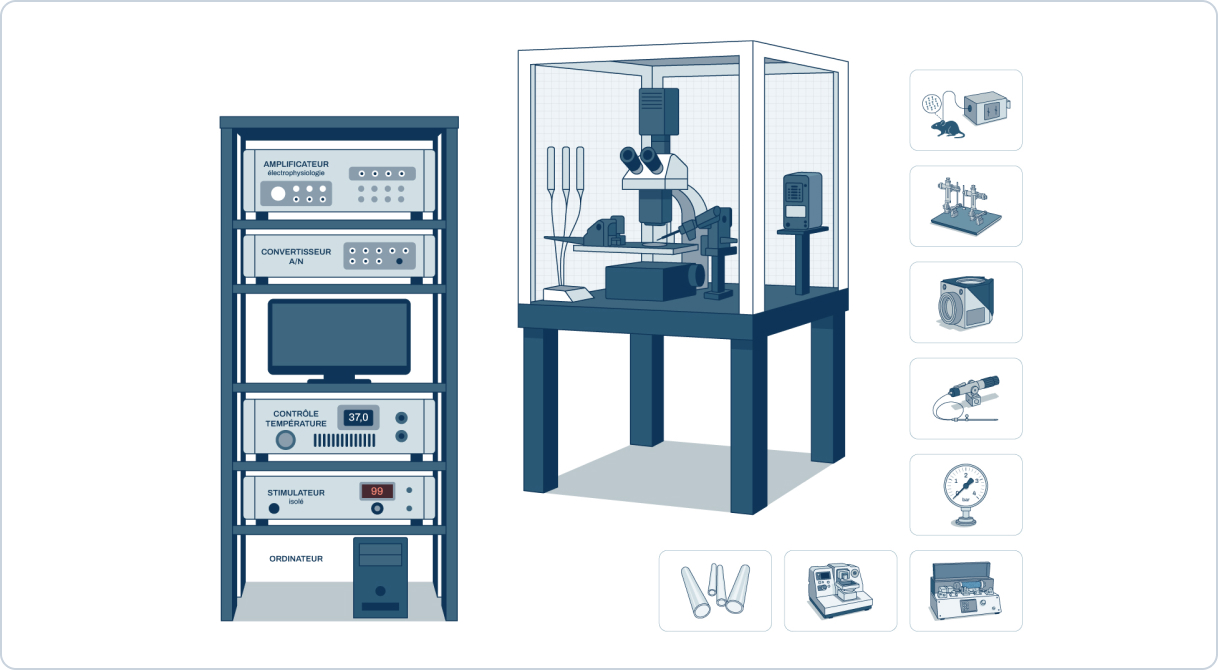
Applications
- Fluorescence imaging of living cells (widefield)
- Phase contrast, Normaski (DIC) and brightfield imaging
- Confocal spinning disk
- FLIM microscopy combined with high-speed intensifier
- Advanced techniques: super-resolution, calcium imaging, FRET, LightSheet, HILO, OPT