

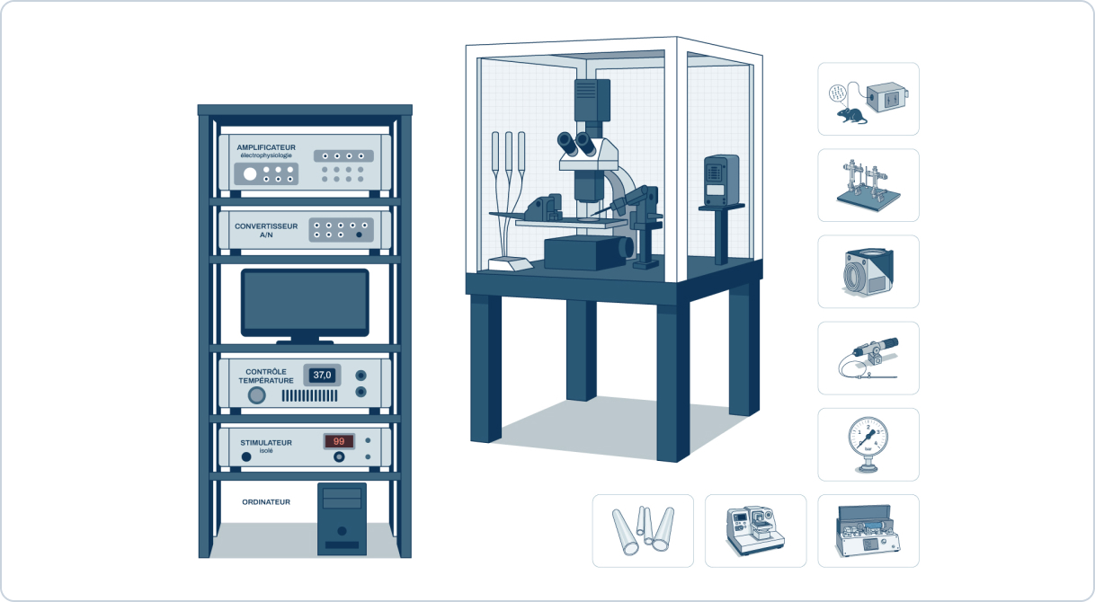
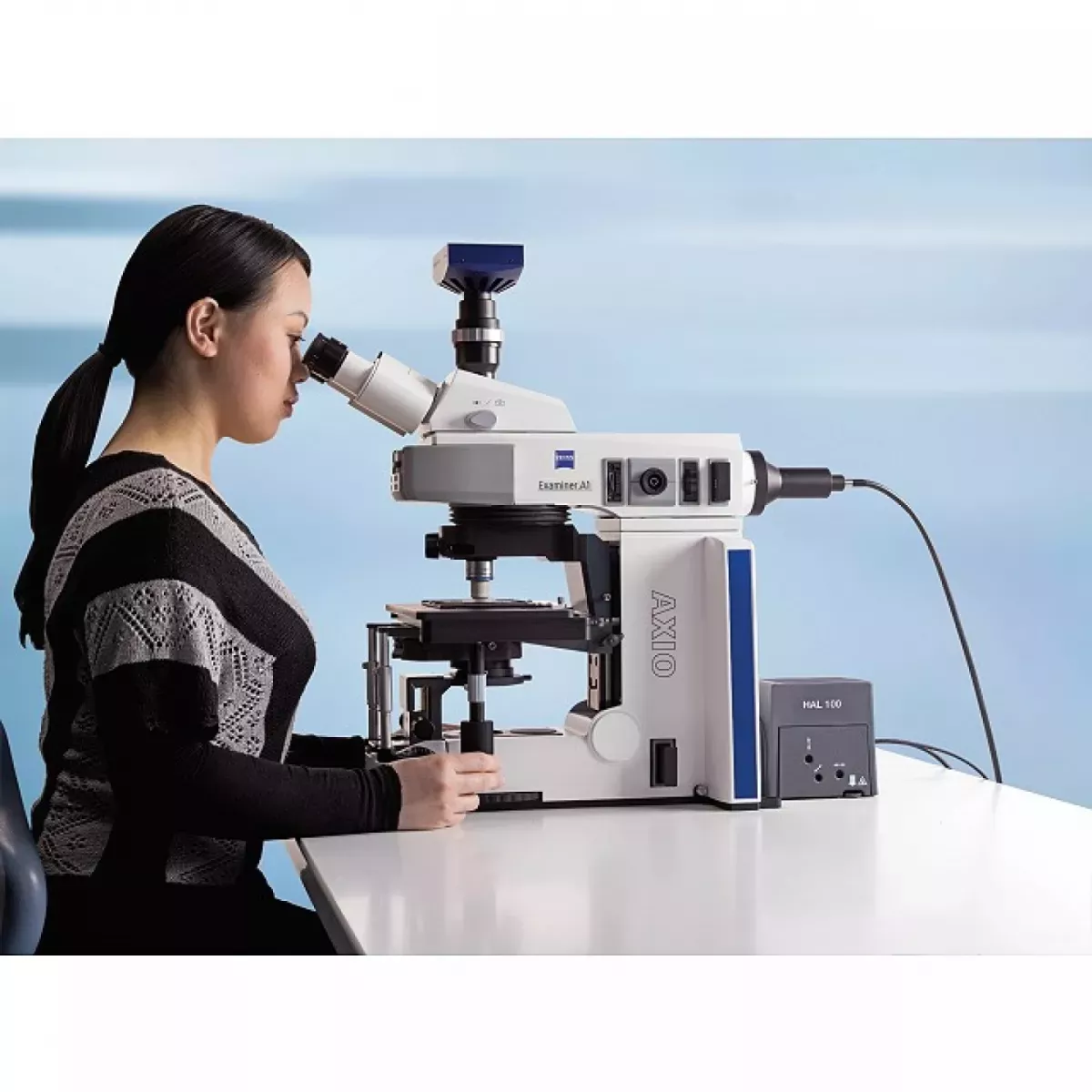
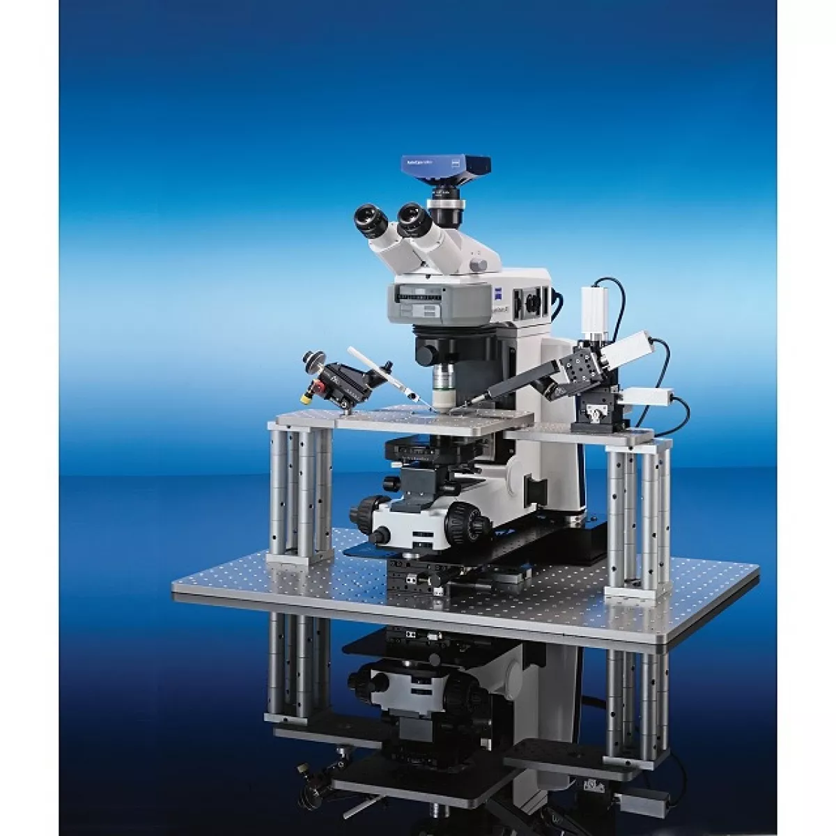
ZEISS AxioExaminer microscope – Configuration 1
The Axio Examiner.A1 Configuration 1 from ZEISS is designed for patch clamp experiments and other applications requiring optimal access to the sample. The microscope maximises the space around the preparation by shifting the optical axis to the front of the objective turret and replacing the objectives by lifting and rotating them backwards, thus avoiding interference with the micro-pipettes.
The stage and condenser can be adjusted independently, providing a working height of over 100 mm. The main adjustments – focus, stage height, condenser diaphragm, dia- and episcopic illumination, light intensity and Dodt gradient contrast – are accessible from the front of the microscope.
The optical system has been specifically developed for electrophysiology and allows several observation techniques to be combined on the same instrument: single oblique observation and Dodt gradient contrast. Simple oblique observation is integrated into the condenser with 360° rotation of the diaphragm, while Dodt gradient contrast offers a cost-effective alternative for observing thick preparations, with a pull-pull polariser (visible light/IR positions) and ‘Push & Click’ analyser.
Two long-distance condensers are available, with numerical apertures of 0.8 and 0.9, for great optical flexibility depending on the application.
Features of the ZEISS AxioExaminer Microscope – Configuration 1
- Optical axis offset towards the front of the turret to maximise working space
- Objectives replaced by lifting (22 mm) then rotating backwards, away from the micro-pipettes
- Stage and condenser independently adjustable
- Working height > 100 mm
- Front-accessible settings: focus, stage, condenser and diaphragm, diascopic/episcopic illumination, transmitted light intensity, Dodt gradient contrast
- Single oblique observation integrated with condenser, 360° pivoting diaphragm
- Dodt gradient contrast observation: 360° pivoting diaphragm, pull polariser (visible/IR), Push & Click analyser
- Long-distance condensers: 0.8 and 0.9 numerical aperture
- Optical and imaging systems adapted to electrophysiology
- Possibility of combining several observation techniques on the same microscope
Documentation
Brochure
PDF file – 1298.6 ko