

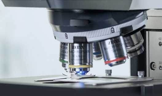
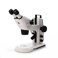
Stemi 305 Stereomicroscope – Zeiss
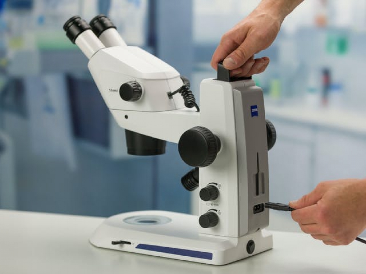
The Stemi 305 is a versatile stereomicroscope, ideal for teaching, quality control, dissection and magnifying. It offers a zoom ratio of 5:1 for magnifications of up to 40x, with a comfortable working distance of 110 mm. Its optical head can be configured as binocular or trinocular with 10X/23 mm eyepieces.
Available with a range of modular stands, it can be adapted to suit every need:
- Simple K stand: basic version, for use with an external light source.
- K MAT stand (industry): equipped with varied reflected lighting, a segmentable LED ring and ESD protection.
- K EDU stand (education): combining reflected and transmitted light, with transmitted light base and polarisation options, ideal for a digital environment thanks to the Stemi 305 camera and Labscope application.
- K LAB stand (laboratory): designed for advanced observations, including oblique brightfield, with multiple lighting sources (LED spotlights, double gooseneck, ring, tilting mirror).
With its many lighting options and ergonomic design, the Stemi 305 is a high-performance, adaptable solution for educational, industrial and scientific environments.
Features of the Stemi 305 stereomicroscope
The Stemi 305 stereomicroscope has been designed for demanding teaching, quality control, dissection and work under magnifying glass.
- Customisable binocular or trinocular optical head, 10X/23 mm eyepiece
- Zoom ratio of 5:1
With a magnification of up to 40x and a working distance of 110 mm, it is available on a modular K stand in several versions
Simple K stand without integrated electronics for use with an external light source
K MAT stand
- Industry
- Allows a variety of reflected light illuminations (episcopy). This stand is ESD (electrostatic discharge protected).
- Binocular tube
- Near-vertical integrated lighting
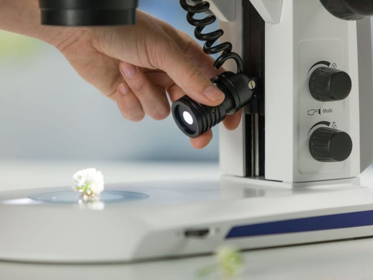
- Segmentable LED ring light for shadow-free ring illumination and oblique light segment illumination: half circle, quarter circle, two spots
- Rotating light segments
- ESD properties: antistatic coating of microscope body and stand
K EDU stand
- Teaching
- Allows varied illumination in reflected light (episcopy) and transmitted light (diascopy)
- Binocular tube
- Integrated, almost vertical lighting
- LED spotlight, zoomable and height-adjustable, for grazing and oblique lighting with cast shadows
- Flat transmitted light base for brightfield and darkfield illumination
- Optional: polarisation equipment for point and transmitted light
- For a digital classroom environment, choose the Stemi 305 cam with K EDU support and K LED spotlight
K LAB stand
- Laboratory
- Allows a variety of illuminations in reflected light (episcopy) and transmitted light (diascopy), in particular the oblique brightfield, essential for observing oocytes and embryos.
- Binocular tube
- Near-vertical integrated lighting
- Self-supporting two-armed gooseneck for variable oblique illumination with distinct shadow effect
- Tilting mirror base for brightfield, darkfield and oblique illumination
- Optional: ergonomic hand rest, polarisation equipment for spotlights and transmitted light
- The wide range of lighting options includes single LED spotlights, self-supporting double spotlights, segmentable ring spotlights and semi-vertical spotlights for cavity lighting.
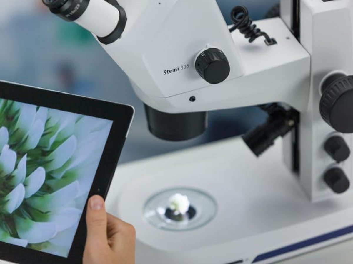
The Labscope iPad imaging app displays live images from all Stemi 305 microscopes in your network on every connected iPad. With a simple tap, you can view each student’s result. It’s easy to capture an image, add annotations and measurements and save it – or export it directly to your server. The fixed 50/50 split is ideal for live demonstrations: your students or customers can follow your microscopic work live on the monitor.
Documentation
Stemi 305 brochure
PDF file – 983.4 ko