

MS-OPC Oocyte Recording Chamber ALA Scientific
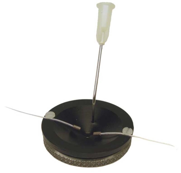
The MS-OPC oocyte perfusion chamber is a device designed to efficiently channel perfusion solutions around an oocyte.
The oocyte is positioned at the bottom of a funnel, on a glass slide, where it is well bathed by the injection of solution via a metal cannula.
The funnel shape means that the volume at the bottom is minimal, guaranteeing very rapid exchange of solution while providing sufficient lateral space for the recording electrodes.
Features of the Oocyte Perfusion Chamber (MS-OPC)
- Funnel shape to optimise oocyte contact with the solution, ensuring very rapid and homogeneous exchange
- Positioning of the oocyte on the glass slide at the bottom of the cone for optimal bathing and electrical stability
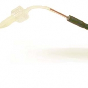
- 19-gage infusion cannula supplied, usable for perfusion or aspiration depending on assembly (with or without Levelock™ to manage overflow)
- Reduced working volume for almost instantaneous solution change
- Sufficient volume around the cone for placement of two voltage clamp recording electrodes
Technical characteristics of the Oocyte Perfusion Chamber (MS-OPC)
| Caractéristique | Valeur |
|---|---|
| Model | MS-OPC |
| Type of room | Funnel chamber oocyte infusion |
| Useful volume | Very small (~4-20 µl), ensuring rapid infusion |
| Injection point | 19-gage metal cannula (injection at the bottom of the cone) |
| Position of the oocyte | On a glass slide (#1 cover slip) at the bottom of the cone |
| Fluid level management | Funnel effect automatically controls infusion level |
| Compatibility | Compatible with optical supports with side access for electrodes |
| Electrophysiological applications | Use of patch clamp / two electrode voltage clamp on Xenopus oocytes |
| Cleaning / maintenance | Simple assembly, easy removal for sterilisation and cleaning |
Applications
- Electrophysiological recordings on oocytes (Xenopus in particular) via voltage clamp or patch clamp
- Pharmacological studies requiring rapid changes of solution around the oocyte
- Laboratories and automated oocyte recording platforms
- Research or teaching in the physiology and expression of ionic receptors
Documentation
ALA Scientific MS-OPC Manual
PDF file – 464 ko