

INFINITY5-5M monochrome USB3 HDMI 5M 2/3” camera
Teledyne Lumenera INFINITY5-5M (INFINITY5-5M model)
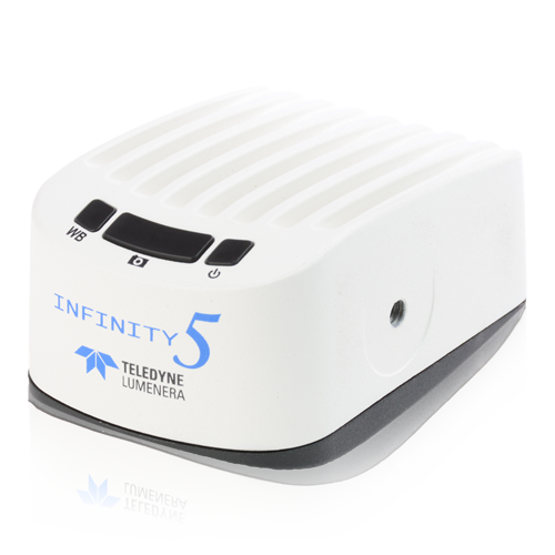
Description of the Teledyne Lumenera INFINITY5-5M camera
5 MP monochrome CMOS digital scientific camera with dual USB 3.1 and HDMI outputs, ideal for advanced fluorescence microscopy, dark field and shared observation.
Based on the Sony IMX250 CMOS sensor with global shutter, it delivers high-resolution images (2464 × 2056) at a speed of 75 fps.
It is ideally suited to fluorescence, DIC or teaching applications, with real-time display and no latency.
Its intuitive design with on-board buttons (power, white balance, capture) makes it easy to use standalone or connected via USB 3.0 to a computer.
Features of the Teledyne Lumenera INFINITY5-5M camera
- Sony IMX250 monochrome CMOS sensor (2/3″) with global shutter
- Resolution 2464 × 2056 pixels (5 MP)
- 3.45 µm pixels for excellent sensitivity
- Playback speed: up to 75 fps in 8 bits
- Dual USB 3.1 Gen 1 and HDMI output for simultaneous viewing
- On-board buttons: power, white balance, image capture
- Playback noise: 2.3 e-
- Saturation capacity: 10,500 e-
- Dynamic range: 72 dB
- INFINITY ANALYZE 7 software included, no annual licence required
Technical specifications of the Teledyne Lumenera INFINITY5-5M camera
| Caractéristique | Valeur |
|---|---|
| Sensor | Sony IMX250, monochrome CMOS |
| Resolution | 2464 × 2056 pixels |
| Pixel size | 3.45 µm |
| Sensor format | 2/3″ (11.1 mm) |
| Saturation capacity | 10 500 e- |
| Playback noise | 2,3 e- |
| Dynamic range | 72 dB |
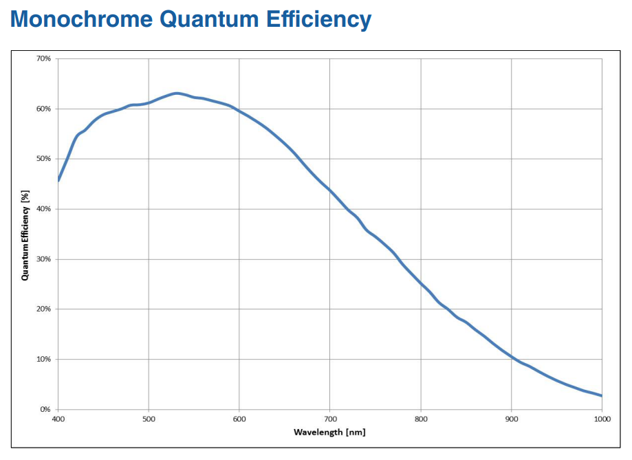
| Quantum efficiency (QE) | 63 % |
| Max. speed | 75 fps |
| Interface | USB 3.0, HDMI |
| Frame | C-mount |
| Weight | 330 g |
| Dimensions | 97.8 × 69.8 × 50.8 mm |
| Power supply | 5 V DC, 1.2 A |
| Operating temperature | 0 to 50 °C |
| Operating humidity | 5 to 95%, non-condensing |
Applications
- Low-light fluorescence
- Immunofluorescence
- Dark field, DIC/Phase
- DNA analysis
- Live cell imaging
- Whole slide imaging
- Histology, pathology, cytology
- Calcium/ionic imaging
- Forensic analysis
- Semiconductor inspection
- Metallurgical microscopy
- Gel documentation
Documentation
Brochure
PDF file – 3427 ko