

INFINITY8-8M monochrome 8.3M 1/1.8” camera
8.3 Megapixel CMOS monochrome digital scientific camera, ideal for standard bright and dark field microscopy techniques, DIC, and general applications.
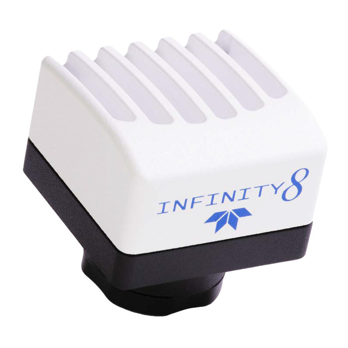
Description of the Teledyne Lumenera INFINITY8-8M camera
Designed with the Sony IMX334 CMOS sensor (1/1.8″ format), it offers a resolution of 3840 × 2160 pixels at a frame rate of 44 frames per second.
It’s perfect for upgrading an existing microscope and easily sharing images or videos.
Although it is not suitable for fluorescence imaging, it excels in bright field, dark field and other applications requiring excellent monochrome rendering.
It comes with INFINITY ANALYZE 7 software at no extra cost, and is compatible with Windows and macOS.
Features of the Teledyne Lumenera INFINITY8-8M Camera
- Sony IMX334 monochrome CMOS sensor (1/1.8″) with rolling shutter
- Resolution 3840 × 2160 pixels (8.3 MP)
- 2 µm pixels for high resolution
- Playback speed: up to 44 fps (full resolution)
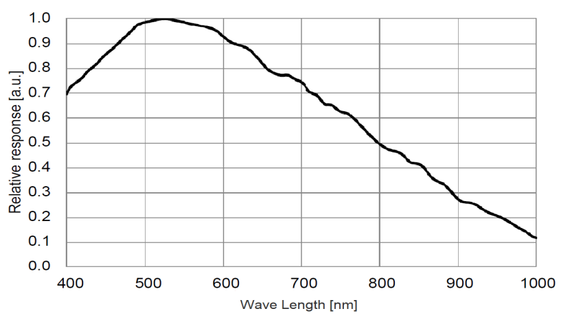
- Quantum efficiency: 82
- Dynamic range: 73 dB
- Saturation capacity: 7030 electrons
- Read noise: 2 e-
- Acquisition: single image, time-lapse, video
- USB 3.0 interface with lockable connector
- INFINITY ANALYZE software included, no annual licence required
Technical specifications of the Teledyne Lumenera INFINITY8-8M camera
| Caractéristique | Valeur |
|---|---|
| Sensor | Sony IMX334, monochrome CMOS |
| Resolution | 3840 × 2160 pixels |
| Pixel size | 2 µm |
| Sensor format | 1/1.8″ |
| Saturation capacity | 7030 e- |
| Playback noise | 2 e- |
| Dynamic range | 73 dB |
| Quantum efficiency (QE) | 82 % |
| Max. speed | 44 fps |
| Interface | USB 3.0 (5 Gbps) |
| Frame | C-mount |
| Weight | 108 g |
| Dimensions | 41.5 × 45 × 45 mm |
| Power supply | USB or 5-25 V via GPIO |
| Operating temperature | 0 to 50 °C |
| Operating humidity | 5 to 95%, non-condensing |
Applications
- Dark field
- DNA analysis
- Live cell imaging
- Whole slide imaging
- Near infrared DIC imaging
- Histology, pathology, cytology
- Calcium/ionic imaging
- Forensic analysis
- Semiconductor inspection
- Metallurgical microscopy
- Gel documentation
Documentation
Brochure
PDF file – 2360.4 ko