

INFINITY8-2M Routine Fluorescence Monochrome 1.7M 1.1” Camera
Teledyne Lumenera INFINITY8-2M (Model I8-LM00-02M)
1.7 Megapixel CMOS monochrome digital scientific camera, optimised for fluorescence imaging, bright field, dark field and other advanced techniques.
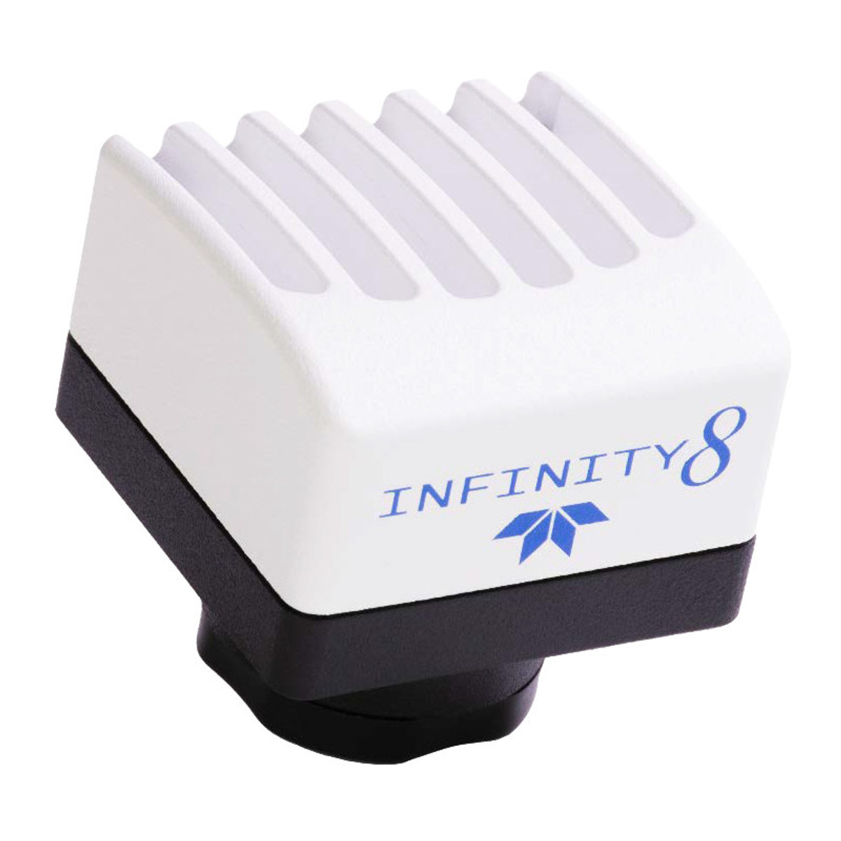
Description of the Teledyne Lumenera INFINITY8-2M camera
Featuring large pixels and a 1.1-inch Sony IMX432 CMOS sensor, this camera offers excellent light sensitivity, ideal for laboratories on a limited budget wishing to image weakly fluorescent samples.
With a frame rate of up to 96 frames per second, a direct USB 3.0 interface and a compact design with passive heat dissipation, it is a high-performance, affordable solution.
Features of the Teledyne Lumenera INFINITY8-2M Camera
- Sony IMX432 1.1″ monochrome CMOS sensor
- 1608 × 1104 pixel resolution (1.7 MP)
- Large 9 µm pixels for increased sensitivity
- Playback speed: up to 96 fps
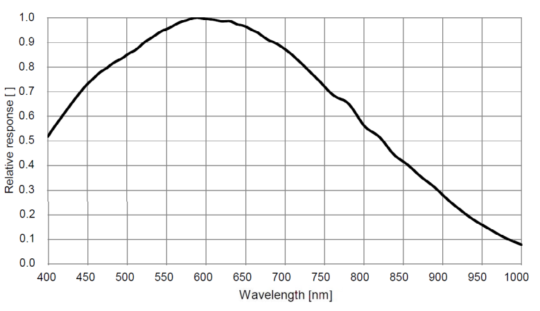
- Quantum efficiency: 76
- Dynamic range: 72 dB
- Saturation capacity: 100,400 electrons
- Low read noise: 4.4 e- (HCG mode)
- Acquisition: single image, fluorescence, time-lapse, video
- Direct USB 3.0 interface (industrial locking)
- INFINITY ANALYZE software included (Windows/Mac, no annual licence required)
Technical specifications of the Teledyne Lumenera INFINITY8-2M camera
| Caractéristique | Valeur |
|---|---|
| Sensor | Sony IMX432, monochrome CMOS |
| Resolution | 1608 × 1104 pixels |
| Pixel size | 9 µm |
| Sensor format | 1.1″ |
| Saturation capacity | 100 400 e- |
| Playback noise | 4.4 e- (HCG mode) |
| Dynamic range | 72 dB |
| Quantum efficiency (QE) | 76 % |
| Max. speed | 96 fps |
| Interface | USB 3.0 (5 Gbps) |
| Frame | C-mount |
| Weight | 108 g |
| Dimensions | 41.5 × 45 × 45 mm |
| Power supply | USB or 5-25 V via GPIO |
| Operating temperature | 0 to 50 °C |
| Operating humidity | 5 to 95%, non-condensing |
Applications
- Low-light fluorescence imaging
- Immunofluorescence
- Bright field, dark field, DIC/phase
- DNA analysis
- Live cell imaging
- Whole slide imaging
- Near infrared DIC imaging
- Histology, pathology, cytology
- Calcium/ionic imaging
- Forensic analysis
- Semiconductor inspection
- Metallurgical microscopy
- Gel documentation
Documentation
Brochure
PDF file – 2360.4 ko