

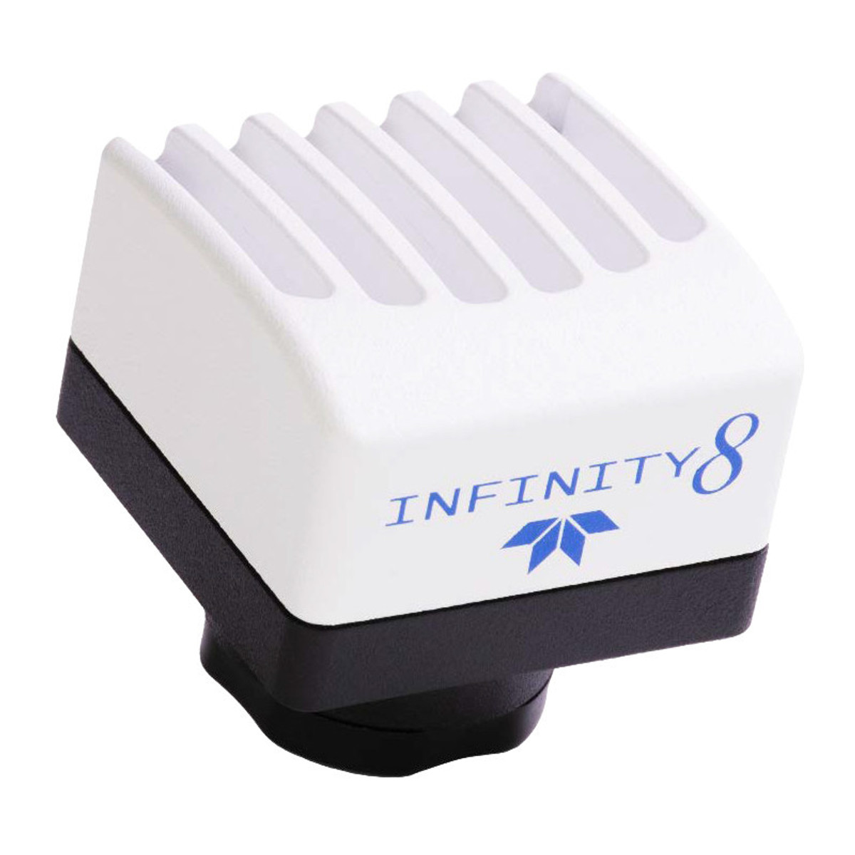
INFINITY8-3M 2.9M 2/3” Monochrome Camera
Teledyne Lumenera INFINITY8-3M (Model I8-LM00-03M)
2.8 Megapixel CMOS monochrome digital scientific camera, optimised for clinical
life sciences and materials science applications.
Description of the Teledyne Lumenera INFINITY8-3M camera
Equipped with a Sony IMX429 CMOS sensor with global shutter and a 2/3″ format, it
captures detailed images at 62 frames per second in full resolution.
With a resolution of 1944 × 1472 pixels and the ability to capture up to 124 fps in sub-sampling mode, this
sub-sampling mode, this camera combines precision, speed and fluidity of display
without latency.
It comes with INFINITY ANALYZE software, free of charge and compatible with Windows
and macOS compatible.
Features of the Teledyne Lumenera INFINITY8-3M camera
- Sony IMX429 (2/3) monochrome CMOS sensor with global shutter
- Resolution 1944 × 1472 pixels (2.8 MP)
- 4.5 µm pixels for high sensitivity
- Playback speed: up to 62 fps (full resolution), 124 fps (sub-sampling)
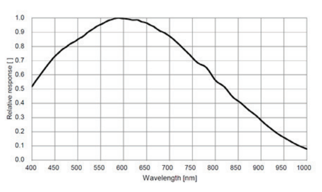
- Quantum efficiency: 74
- Dynamic range: 72 dB
- Saturation capacity: 24,900 electrons
- Read noise: 7.2 e-
- Acquisition: single image, fluorescence, time-lapse, video
- USB 3.0 interface with lockable connector
- INFINITY ANALYZE software included, no annual licence required
Technical specifications of the Teledyne Lumenera INFINITY8-3M camera
| Caractéristique | Valeur |
|---|---|
| Sensor | Sony IMX429, monochrome CMOS |
| Resolution | 1944 × 1472 pixels |
| Pixel size | 4.5 µm |
| Sensor format | 2/3″ |
| Saturation capacity | 24 900 e- |
| Playback noise | 7,2 e- |
| Dynamic range | 72 dB |
| Quantum efficiency (QE) | 74 % |
| Max. speed | 62 fps (1944×1472), 124 fps (968×736) |
| Interface | USB 3.0 (5 Gbps) |
| Frame | C-mount |
| Weight | 108 g |
| Dimensions | 41.5 × 45 × 45 mm |
| Power supply | USB or 5-25 V via GPIO |
| Operating temperature | 0 to 50 °C |
| Operating humidity | 5 to 95%, non-condensing |
Applications
- Low-light fluorescence imaging
- Immunofluorescence
- Dark field, DIC/phase
- DNA analysis
- Live cell imaging
- Whole slide imaging
- Near infrared DIC imaging
- Histology, pathology, cytology
- Calcium/ionic imaging
- Forensic analysis
- Semiconductor inspection
- Metallurgical microscopy
- Gel documentation
Documentation
Brochure
PDF file – 2360.4 ko