

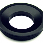
ALA Scientific round chamber Ø 25mm MS-502 ALA Scientific
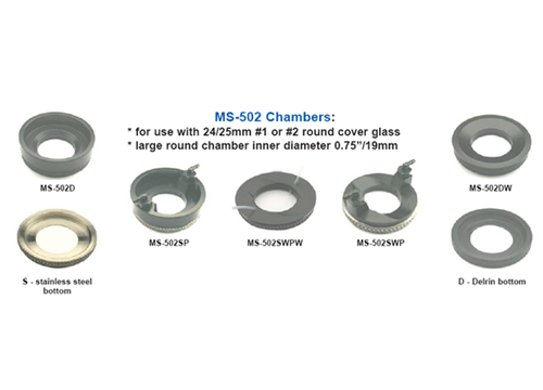
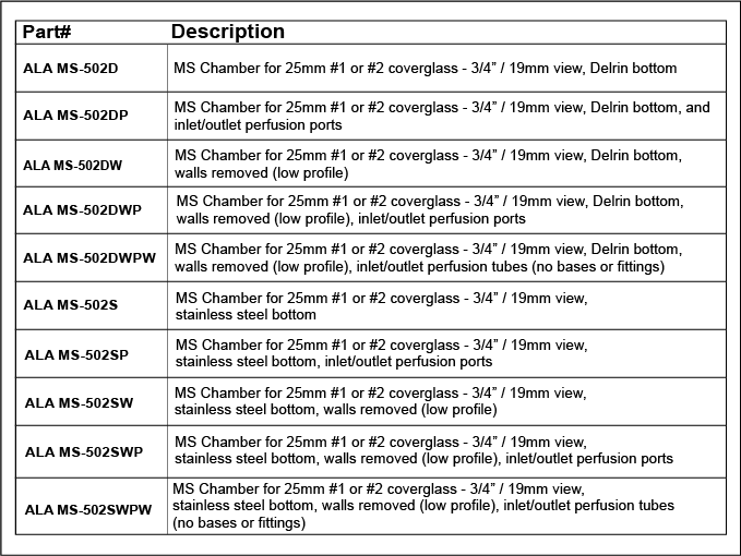
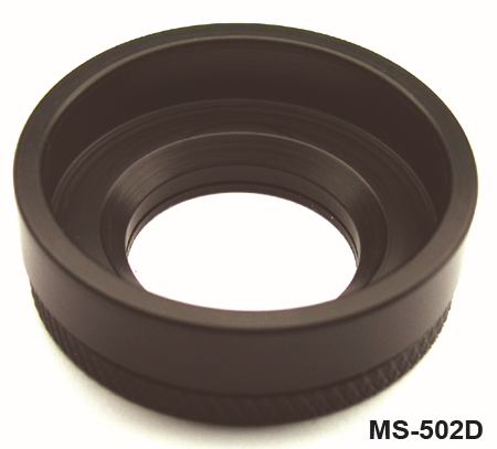
The MS-502 round chamber from the MS series is designed to secure tissue preparations or cells on 25 mm round cover glass (#1 or #2). It is compatible with specimen holders such as MS-Stage, HCMIS and 35 mm Petri plates, ensuring stable mounting without glue or grease thanks to its two compressible O-rings.
Features of the ALA Scientific Ø 25mm MS-502 Round Chamber
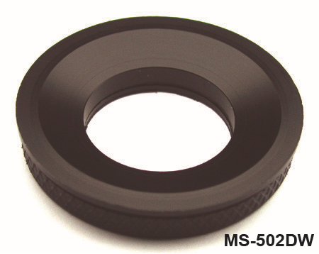
- Round inner diameter of 0.75″ (19 mm) exactly adapted to 25 mm glass
- Double O-ring seal (tightness and retention)
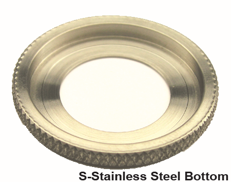
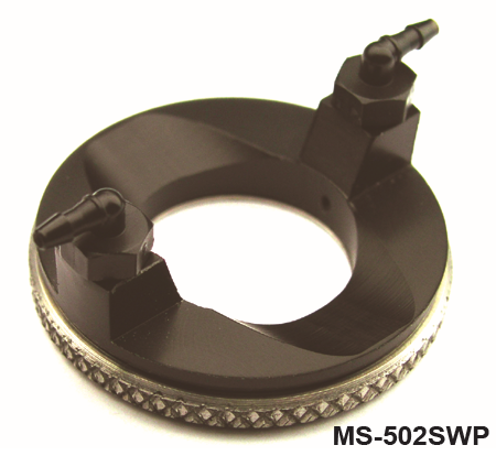
- Interchangeable backs: stainless steel for thermal control or black Delrin for imaging
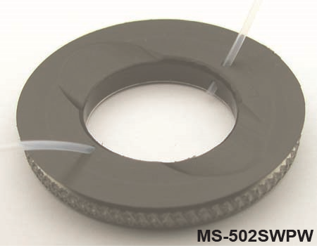
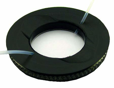
- Low mount or removable walls for electrode access
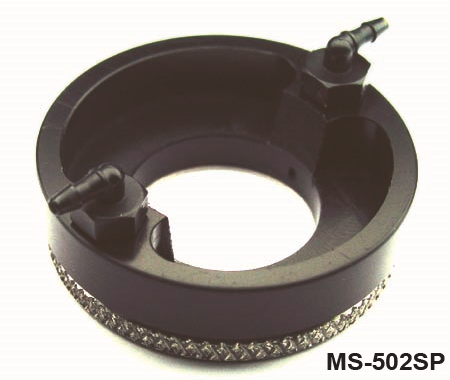
- Integrated perfusion options (1/16″ ports)
- Thermal foil option (T), always with stainless steel base
Technical characteristics of the ALA Scientific Round Chamber Ø 25mm MS-502
| Caractéristique | Valeur |
|---|---|
| Type of room | MS-502 (round for glass covering 24-25 mm #1/#2) |
| Inside diameter | 0.75″ / 19 mm |
| Bottom materials | 316 SS stainless steel or black Delrin |
| O-rings | Double compression seals |
| Walls | Normal, low or removable (W) |
| Infusion ports | Optional P = 2 ports 1/16″, with or without walls (W) |
| Thermal option (foil) | Foil T, stainless steel bottom compulsory |
| Compatibility | MS-Stage, 35 mm Petri, HCMIS |
| Volume of solution | Low (low profile, laminar flow) |
| Recommended use | Electrophysiology, cellular imaging, perfusion |
Applications
- Electrophysiology experiments (patch clamp, recordings)
- Fluorescence imaging on cells/tissues close to the surface
- Controlled perfusion (laminar flow) in stable, sterile conditions
Documentation
Brochure
PDF file – 274.2 ko